Research Studies
Research Studies at The MR Research Centre
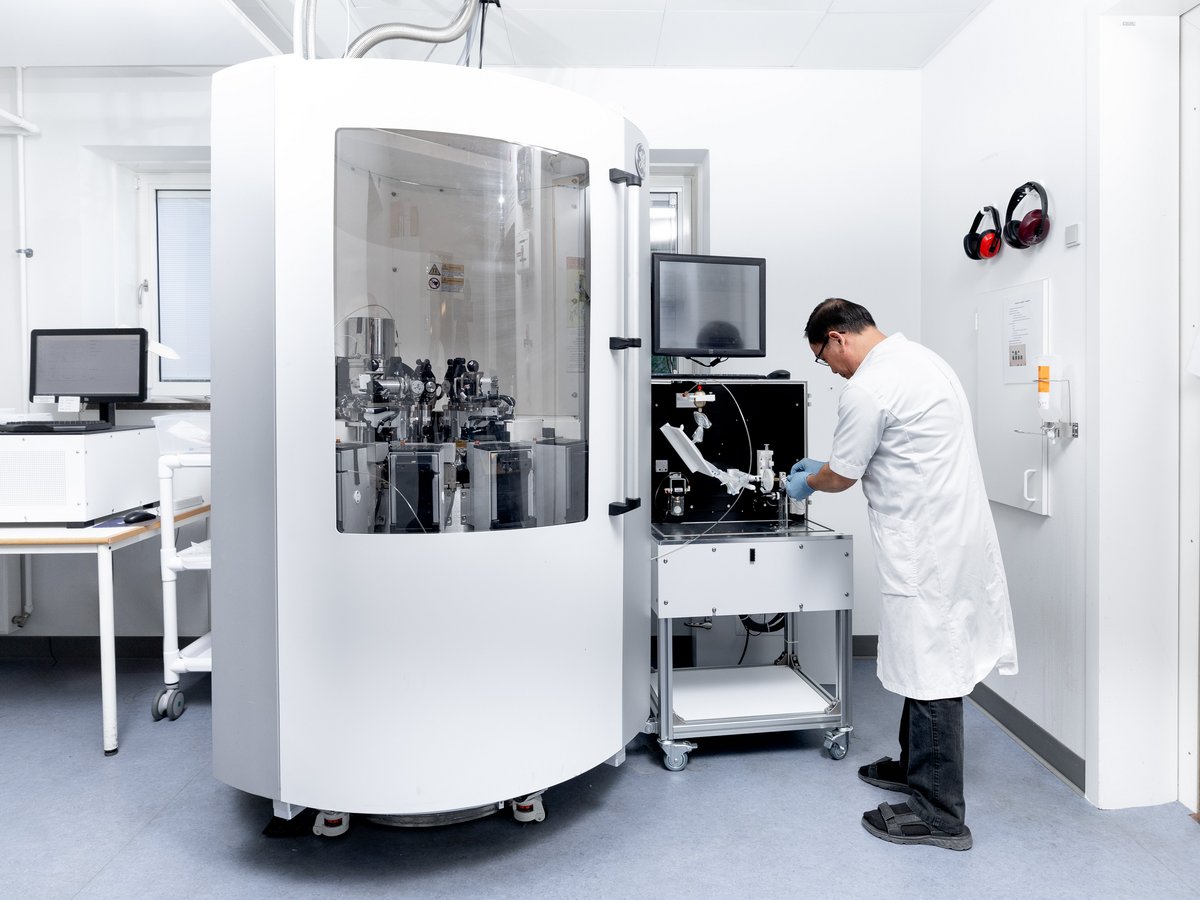
At the MR Research Centre at Aarhus University, we develop the methods below to bring them to clinical use. We make unique recordings of the body's physiology using MRI scanning employing carbon, phosphorus, sodium, xenon, and deuterium.
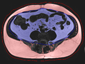
Clinical MR scanners are powerful superconducting magnets that can create images of the inside of the body using harmless radio waves. The method allows signal recording from soft tissue and can distinguish tiny changes in tissue types, making it unique.
You usually see images of hydrogen atoms bound in water and fat.
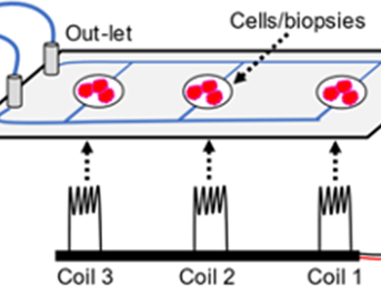
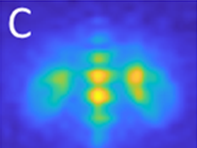
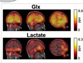
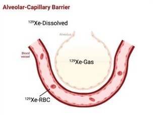
Still, the MR scanner can do much more: It records signals from magnetic nuclei of other elements such as carbon (13C), sodium (23Na), phosphorus (31P), deuterium (2H), and xenon (129Xe). This technology, known as multinuclear spectroscopy (MNS), has excellent clinical and research potential in imaging specific molecules and essential metabolic functions.
At the MR Centre, Aarhus University, we use MNS daily, and we are working on fully developing them for clinical use to improve the clinical applicability of MR scans.